
 |
The Montreal Neurological Institute (MNI) is a teaching and research institute of McGill University , founded by Wilder Penfield in 1934. His vision was to bring basic research scientists and clinicians together in one institute where all could work towards a common goal of understanding the brain. Such proximity promotes exchange and enriches both the understanding and the treatment of neurological diseases. We use several approaches in our studies of human brain function, and usually we combine approaches. Some of our studies begin with pilot studies carried out with college students, to establish how a particular neuropsychological task is performed by a healthy brain. Once satisfied that a task is appropriate for answering a particular question, we use it to test patients with brain lesions or with epileptic abnormalities, or with surgical resections in known sites. This provides information about the effect of brain damage in specific sites on the function tapped by the task. We then test healthy volunteers on the task in a functional neuroimaging paradigm; this provides information about what brain regions are important for performing the task. |
|
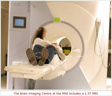
Finally, with certain tasks we also perform the functional neuroimaging test with patients. If they are able to perform the task, the neuroimaging results tells us how they are able to compensate for the damaged areas of the brain. These findings together provide a rich picture of how the brain functions for a given task. The results also help us design neuropsychological tests that we can expect to be sensitive to dysfunction in specific areas of the brain. These tests are useful for diagnosis in patients whose site of damage is not known, and also for following patients over time or after surgical intervention to determine whether the patient shows losses or improvements (or remains stable). We use positron emission tomography (PET) or functional magnetic resonance imaging (fMRI) for our functional neuroimaging studies, depending on the research question. These studies are carried out in the McConnell Brain Imaging Center (BIC), where we have access to high technology brain scanners and sophisticated computational analyses. Shown in the photograph is the 1.5T scanner. The lab's research focuses on three main areas: chemosensory, epilepsy, and memory research. In each of these the ultimate goal is to elucidate neuronal substrates behind well defined behavioral responses, or phenomena, with the broader aim of reaching a greater understanding of the human brain. Among projects that are currently in progress are: |
|
|
|
||
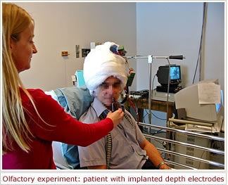
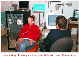
Olfaction, emotions, and memories . One line of our research relates to the innate emotionality of odors. In this project we are investigating the behavioral and neuronal responses to emotionally arousing odors. Among other measures, olfactory and gustatory evoked potentials are examined in patients with depth electrodes implanted in the amygdala and hippocampus, and behavioral responses are studied in patients with a unilateral temporal-lobe resection. Chemosensory communication . Whether humans use chemosignals as a mean of non-conscious communication is a debated question. We are currently investigating different putative human pheromonal compounds in behavioral and neuroimaging studies. Olfactory perception . To date, our knowledge of the olfactory sense is derived mostly from perception of single, pure odorants. However, most odors that we perceive in our daily life are not pure odorants, but consist of mixtures of different odorants and compounds with a trigeminal component. In one series of experiments we are investigating the neuronal substrate of the olfactory perception of everyday odors using measures ranging from receptor activation (negative mucosa potentials) to central activation (studied with EEG, PET and fMRI) to behavior. This work is performed in collaboration with Mats Olsson and Thomas Hummel . |
||
|
|
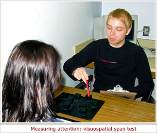
Memory and temporal-lobe function . The relative roles of novelty/familiarity versus verbal/nonverbal stimuli in learning and retention are not well known. In these experiments, we aim to disentangle these different factors by investigating the neuronal substrate in patients with left or right medial temporal-lobe dysfunction and in healthy individuals using fMRI and behavioral measurements. Attention . In this project, we are teasing out the critical variables in a pair of matched verbal versus visuospatial attention tasks and determining the extent to which these are sensitive to lateralized temporal-lobe dysfunction. Intracarotid anesthetic procedures (IAPs) . The intracarotid amobarbital procedure is an important part of a comprehensive investigation of patients who are candidates for surgical treatment of epilepsy. Owing to frequent and lengthy shortages of sodium amobarbital, we have initiated the use of a different anesthetic agent (etomidate), and with it we introduced a significant change in the procedure, such that now the time constraints are essentially abolished. We are also working towards development of a reliable fMRI protocol that will yield information currently sought in the IAP memory test. Neuropsychological tests . Part of our work relates to development of or improvement of clinical neuropsychological tests. In our specialized setting, patients are usually tested more than once, such as before and after surgery, or at two or more intervals when a deterioration is suspected. In addition, a large proportion of our patients are francophone (and the others, anglophone). Therefore we develop our tests in equivalent alternate forms, to be used in retesting, and in both languages. Examples of our newer work include our test for subtle parietal-lobe dysfunction using haptic search, in collaboration with Viviane Sziklas and Marta Maximov, our test of right-hemisphere visuoperceptual functioning, and a test of social perception that we expect to be sensitive to frontal-lobe dysfunction. |
|
|
||
and The Savoy Foundation. |
||

Montreal Neurological Institute
McGill University
© 2005